By Dr. Gabriel Rodriguez
Introduction
Mindfulness meditation has exploded in popularity over the last few decades, becoming a widespread therapeutic technique used to reduce stress, anxiety, depression and pain. But beyond the hype, growing research using neuroimaging technologies reveals that the consistent practice of mindfulness meditation may actually change the physical structure and wiring of the brain over time. In ways both ephemeral and enduring, mindfulness sculpts the mind by molding the brain.
Brain Regions Activated During Meditation
Functional magnetic resonance imaging (fMRI) studies show that the practice of mindfulness meditation activates a widespread network of brain regions involved in executive control, emotional regulation, meta-awareness, and attentional processing.
- The Prefrontal Cortex and Anterior Cingulate Cortex
Key nodes in this network include the prefrontal cortex (PFC) and anterior cingulate cortex (ACC). The PFC plays a major role in executive functions like planning, decision-making, and modulating emotions. Specifically, mindfulness meditation enhances activity in the dorsolateral PFC, which supports sustained attention and working memory. The ACC is involved in self-regulation, including monitoring emotions and controlling impulses. It also contributes to cognitive flexibility.
During meditation, increased PFC and ACC activation is believed to underlie the meta-cognitive aspects of mindfulness - the ability to monitor mental experiences with clarity and objectivity. The heightened PFC-ACC circuitry allows meditators to notice thoughts, emotions and sensations without attaching narrative judgments to them.
Another structure implicated is the insula, which detects internal body sensations and modulates emotional awareness. The insula has extensive connections to other brain areas involved in attention, perception and emotion. Enhanced insula activation during mindfulness meditation is thought to support a dispassionate, observer-like perspective on transient thoughts, feelings and urges as they arise and pass away.
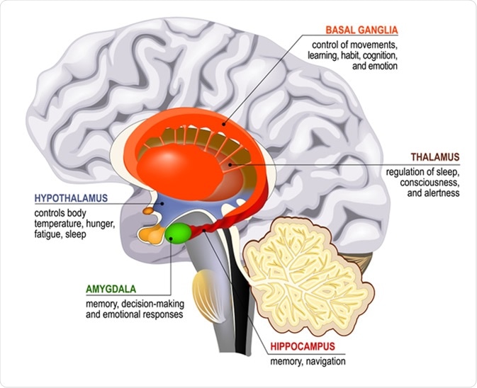
Increased activation of the hippocampus has also been observed during mindfulness practice. The hippocampus plays a key role in learning, context and memory. Along with boosting present-moment awareness, mindfulness meditation may strengthen abilities like associative thinking and memory formation that rely on the hippocampus.
The amygdala is also recruited during meditation, but in a different way than the aforementioned regions. The amygdala is strongly involved in threat detection, fear conditioning and emotional reactivity. While mindfulness practice activates executive control regions like the PFC and ACC, it deactivates the amygdala. This neural decoupling of cognitive control and emotional reactivity regions during meditation may lead to reduced stress and more skillful responses to challenging situations.
Changes in Brain Structure
Emerging research suggests that beyond acutely activating brain networks during practice, mindfulness meditation may also change the brain’s physical structure and morphology when practiced over long timescales.
- Increased Cortical Thickness
Neuroimaging studies reveal that experienced mindfulness meditation practitioners exhibit increased cortical thickness in several regions compared to non-meditators. Specifically, significant increases in gray matter thickness tied to meditation experience are found in the prefrontal cortex (PFC) and right anterior insula.
These changes likely reflect use-dependent cortical plasticity induced by repeated meditation. The thickening may result from increased synaptic connectivity and neural branching triggered by the sustained activation of attentional and salience detection circuits during mindfulness practice.
Along with cortical thickening, brain volume changes have been observed in long-term meditators across multiple studies. The hippocampus, temporoparietal junction, orbitofrontal cortex and cerebellum show increased gray matter volume, while the amygdala displays decreased volume.
This aligns with the functional shifts meditation is thought to produce - growth in brain structures that support body awareness, introspection and emotional balance, coupled with shrinkage in the fear-center amygdala. The brain seems to be remodeled by meditation to reinforce positive traits.
Changes in Functional Connectivity
Beyond changing the brain’s anatomical landscape, mindfulness meditation also appears to rewire the functional connections between different brain regions. This has been witnessed in the “resting state” neural dynamics detected by fMRI scans of experienced meditators.
The default mode network (DMN) is a set of brain regions that activate when the brain is at rest and not focused on an external task. Mindfulness meditators show differences in functional connectivity between two hubs of the DMN - the medial prefrontal cortex and posterior cingulate cortex.
This decoupling is believed to allow meditators to monitor inner experiences with greater objectivity and less self-referential thought. Disconnecting the DMN may be one route mindfulness weakens rumination and strengthens attentional control.
- Strengthening the Salience Network
In contrast, meditators display strengthened connectivity within the salience network - a group of brain regions involved in detecting sensations, thoughts and feelings. Regions like the anterior insula and anterior cingulate cortex exhibit greater coordination at rest.
This reflects meditation enhancing present-moment sensory awareness and introspective abilities. Fortifying salience network connections may account for meditators' increased capacity for mindfulness in daily life.
Changes in Neural Oscillations
Shifting from brain anatomy to brain waves, electroencephalogram (EEG) studies reveal mindfulness meditation also sculpts neural oscillations - the rhythmic electrical firing patterns that reflect varying modes of brain function.
- Increased Alpha and Theta Waves
EEG recordings show mindfulness meditation increases alpha and theta wave activity compared to rest. Alpha waves (8-12 Hz) indicate tranquil, relaxed wakefulness, while theta waves (4-8 Hz) signify inward-focused thinking.
Thus, elevated alpha and theta oscillations likely underpin meditators’ calm, meditative awareness and ability to detach from distracting thoughts and emotions. This brain wave signature represents a blissful, tranquil, introspective state of consciousness.
- Gamma Waves - Heightened Conscious Processing
Intriguingly, some studies also report increased gamma wave activity (25-100 Hz) during meditation, specifically in experienced practitioners. Gamma waves are linked to conscious perception, problem-solving and memory formation.
Heightened gamma oscillations may reflect mindfulness strengthening practitioners’ clarity, precision and richness of moment-to-moment experience. However, more research is needed on gamma waves and meditation.
Changes in Amygdala Reactivity
The amygdala is a brain structure strongly linked to emotional reactivity, particularly negative emotions like fear and anxiety. As discussed earlier, mindfulness meditation helps deactivate the amygdala. With prolonged practice, these state changes can mature into longer-lasting trait alterations that curtail amygdala reactivity.
- Reduced Response to Negative Stimuli
Neuroimaging studies confirm that experienced meditators exhibit reduced amygdala response to negative emotional images compared to novices. The degree of amygdala modulation corresponds to the number of meditation hours practiced.
Long-term meditators also show decreased gray matter volume in the basolateral amygdala compared to non-meditators. Together with reduced reactivity, amygdala downsizing suggests meditation helps regulate negative emotions.
- Correlation with Emotional Stability
Importantly, dampened amygdala activation correlates with lower anxiety and neuroticism and greater emotional stability in meditators, highlighting the behavioral relevance of mindfulness reshaping this emotional brain region.
Conclusion
The growing body of neuroscientific research reviewed here demonstrates that mindfulness meditation not only acutely activates brain networks involved in attention, executive control and emotion regulation during practice, but with sustained training may impart lasting positive changes in brain structure and function that mediate greater emotional balance and attentional mastery.
Key takeaways include that mindfulness practice appears to:
- Increase cortical thickness and volume in prefrontal regions that govern executive functions like self-awareness, focus and impulse control.
- Alter functional connections between default mode and salience network hubs, enhancing sensory clarity and introspection.
- Shift neural oscillations into relaxed yet focused alpha/theta rhythms, along with possible gamma wave enhancement among adept practitioners.
- Produce trait-level decreases in amygdala reactivity and volume, reflecting improved emotional regulation.
In sum, by mobilizing neuroplasticity processes to reinforce attention, introspection, clarity and equanimity, mindfulness meditation may impart enduring beneficial changes upon both mind and brain. However, more research is still needed to further elucidate the specific neural mechanisms of how mindfulness transforms consciousness over time.
Though early in the field’s development, these tantalizing clues suggest mindfulness practice holds remarkable potential to sculpt our neural makeup in an uplifting way. With its roots in ancient contemplative traditions, meditation seems capable of generating positive ripples of change that spread across the interconnected layers of our nervous system, making us not only calmer but sharper, nimbler, and wiser within. Mindfulness may teach us we have far more power to shape our brains than we realize. The future of mindfulness neuroscience looks bright and full of promise, both for unveiling the mysteries of mind-brain connections and discovering meditative methods to unlock our hidden potentials.


